
FAQ for beginners
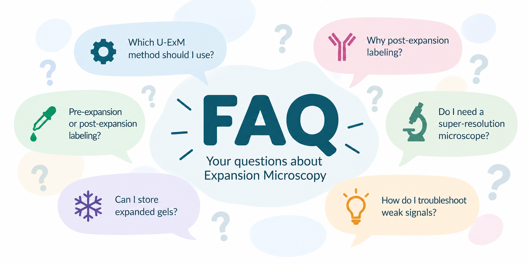
Expansion Microscopy FAQ
Getting started with U-ExM, cryo-ExM and iU-ExM
New to Expansion Microscopy? Here we answer frequently asked questions about choosing a method, preparing samples, immunolabeling, imaging and troubleshooting. These recommendations are based on our laboratory's experience developing and applying ultrastructure-preserving expansion microscopy approaches.
For detailed experimental instructions, visit our Expansion Microscopy Protocols page.
1. Choosing the right ExM method
What is Ultrastructure Expansion Microscopy (U-ExM)?
U-ExM is an expansion microscopy approach designed to preserve the nanoscale organization of cellular structures while physically enlarging the specimen.
Samples are embedded in a swellable polymer network, processed to allow expansion, and expanded in water. This separates molecular structures spatially, allowing features that were previously too close to resolve to be visualized using conventional fluorescence microscopy.
U-ExM typically achieves approximately 4–4.5× linear expansion, providing an effective lateral optical resolution of approximately 60–70 nm with a conventional diffraction-limited microscope, depending on the imaging system.
It is particularly useful for studying centrioles, cilia, microtubules, and other complex cellular structures.
How is U-ExM different from conventional ExM?
Both approaches use swellable polymer gels to physically enlarge biological specimens.
U-ExM was specifically developed and optimized to preserve cellular ultrastructure, with particular attention to structures such as centrioles and microtubules.
Conventional ExM encompasses many different protocols, with varying fixation, anchoring, homogenization and labeling strategies. Ultrastructural preservation therefore depends on the particular protocol and sample.
The important distinction is not simply the expansion factor, but how faithfully the original molecular organization is maintained.
What is cryo-ExM, and why use cryofixation?
Cryo-ExM combines rapid cryofixation with expansion microscopy.
Instead of relying exclusively on chemical fixation, samples are rapidly frozen to immobilize cellular structures before subsequent processing and expansion.
Cryofixation can reduce certain fixation-induced artifacts and improve the initial preservation of sensitive cellular structures.
However, the final result also depends on subsequent sample processing, including freeze substitution, gel embedding and denaturation.
Cryo-ExM does not mean that fluorescence imaging is performed at cryogenic temperature. The specimens are cryofixed during preparation and subsequently expanded for fluorescence imaging.
What is iterative U-ExM (iU-ExM)?
iU-ExM is an iterative expansion approach that uses successive rounds of gel embedding and expansion to achieve substantially higher expansion factors.
Our iU-ExM method typically achieves approximately 16–20× linear expansion, enabling finer structural details to be visualized using fluorescence microscopy.
However, higher expansion factors introduce additional challenges, including sample fragility, fluorescence signal dilution and the need to validate structural preservation carefully.
Which expansion microscopy method should I choose?
The choice depends on the biological sample, the structural question and the available equipment.
| Method | Typical expansion | Main advantage |
|---|---|---|
| U-ExM | ~4–4.5× | Accessible ultrastructure-preserving expansion |
| Cryo-ExM | ~4×, protocol-dependent | Cryofixation-based sample preservation |
| iU-ExM | ~16–20× | Higher effective spatial resolution |
2. Labeling strategies and antibodies
What is the difference between pre-expansion and post-expansion labeling?
In pre-expansion labeling, antibodies or other fluorescent probes are applied before the specimen is embedded and expanded.
In post-expansion labeling, the sample is first processed and expanded, and antibodies are applied afterward.
Both strategies have advantages and limitations. The best choice depends on the target protein, epitope accessibility, sample preparation and the expansion protocol.
Why do we favor post-expansion labeling in U-ExM?
Post-expansion labeling offers several important advantages:
- Improved epitope accessibility: Expansion physically separates cellular components, potentially exposing epitopes that were previously sterically inaccessible.
- Reduced antibody crowding: Dense molecular assemblies become physically enlarged, making it easier for antibodies to access neighboring targets.
- Reduced effective linkage error: Antibodies introduced after expansion are not themselves expanded. Their physical dimensions therefore correspond to smaller distances when expressed in the original biological coordinates.
- Greater labeling flexibility: Expanded samples can be tested with different antibody combinations and staining conditions.
However, post-expansion labeling is not always superior: some epitopes may be modified or lost during processing.
Does post-expansion labeling eliminate antibody linkage error?
No, but it can substantially reduce the contribution of antibody size to localization uncertainty when measurements are expressed in pre-expansion coordinates.
For example, if an antibody introduces a 12 nm displacement after 4× expansion, that displacement corresponds to approximately 3 nm in the original specimen.
By contrast, a displacement introduced before expansion is enlarged together with the sample and is not automatically reduced in pre-expansion coordinates.
This is particularly important when precisely mapping proteins within densely packed molecular structures.
Is post-expansion labeling always better than pre-expansion labeling?
No. Pre-expansion labeling can be advantageous when epitopes are sensitive to denaturation or other expansion-processing steps.
It may also be useful for particular fluorescent proteins, chemical probes or labeling strategies that are designed to retain fluorescence during expansion.
However, pre-expansion labeling requires careful consideration of probe retention, antibody size, steric hindrance and possible displacement of the detected signal.
For challenging targets, we recommend comparing both strategies experimentally whenever feasible.
Can I use antibodies validated for conventional immunofluorescence?
Often, yes, but compatibility is not guaranteed.
An antibody that performs well in conventional immunofluorescence may fail in U-ExM because the epitope is modified during denaturation or other sample-processing steps.
Conversely, some antibodies perform better after expansion because previously inaccessible epitopes become exposed.
We recommend starting with antibodies already validated for U-ExM and optimizing staining conditions when necessary.
Visit our antibody compatibility database for antibodies tested in our laboratory.
Can I perform multicolor immunolabeling after expansion?
Yes. Multicolor labeling is one of the strengths of U-ExM.
As in conventional immunofluorescence, antibody species, secondary antibody cross-reactivity, fluorophore spectra and staining conditions should be considered.
For precise nanoscale measurements, chromatic registration between fluorescence channels is particularly important.
3. Sample preparation and structural preservation
Do I need specialized equipment to perform U-ExM?
Not necessarily. Standard U-ExM can be implemented using equipment commonly available in cell biology laboratories.
Most preparation steps require standard laboratory materials, appropriate chemical reagents, incubators or heating equipment, and access to a fluorescence microscope.
Cryo-ExM additionally requires suitable rapid-freezing equipment, such as a manual plunge freezer or an automated freezing system, depending on the sample.
iU-ExM requires additional gel preparation and handling steps but does not necessarily require specialized imaging hardware.
Why does U-ExM use denaturation rather than protease digestion?
A central objective of U-ExM is to preserve the spatial organization of cellular proteins while allowing the specimen to expand.
Instead of relying on extensive proteolytic digestion, U-ExM uses controlled denaturation to homogenize the specimen mechanically and allow expansion.
This approach can help retain protein-derived structural information and support subsequent immunolabeling.
However, denaturation can alter protein epitopes, and preservation depends on the specific target and processing conditions.
Can I apply U-ExM to any biological sample?
U-ExM has been successfully adapted to a broad range of specimens, including mammalian cells, microorganisms, parasites, cilia and various tissues.
However, no single protocol works optimally for every sample.
Cell walls, extracellular matrices, sample thickness, fixation sensitivity and reagent penetration can all influence expansion.
Some biological systems require adaptations such as additional permeabilization, cell wall treatment or modified sample preparation.
See our Developments and Applications page for examples.
Does cryofixation preserve all cellular structures and epitopes?
No. Cryofixation can provide excellent initial structural preservation by rapidly immobilizing cellular components, but it does not guarantee that every molecular feature remains unchanged.
Subsequent processing steps, including freeze substitution, embedding, denaturation and expansion, may affect membrane organization, protein conformation and epitope recognition.
Therefore, cryo-ExM results should be validated using appropriate structural references and complementary methods whenever possible.
Is expansion isotropic, and how can I verify it?
Ideally, expansion should be isotropic, meaning that structures expand equally in all directions.
In practice, isotropy depends on the specimen, protocol and spatial scale being examined.
A sample can appear globally isotropic while still displaying local distortions.
We recommend validating expansion using well-characterized biological structures, pre- and post-expansion comparisons when possible, or independent structural measurements.
For quantitative studies, the expansion factor should be determined experimentally.
4. Imaging and effective resolution
Do I need a super-resolution microscope for U-ExM?
No. One of the main advantages of expansion microscopy is that nanoscale structural information can be obtained using conventional fluorescence microscopes.
Widefield and confocal microscopy can both be used, depending on sample thickness, fluorescence signal and the biological question.
Combining expansion with techniques such as SIM or STED can further improve effective resolution, provided that labeling density, signal quality and sample stability are sufficient.
How do I calculate effective resolution after expansion?
A commonly used approximation is:
Effective optical resolution = microscope resolution / linear expansion factor
For a microscope with a lateral resolution of 250 nm:
- 4× expansion: approximately 62.5 nm.
- 10× expansion: approximately 25 nm.
- 20× expansion: approximately 12.5 nm.
These values represent nominal optical resolution, not necessarily the actual biological resolution.
Labeling density, antibody geometry, fluorescence signal, structural preservation and optical performance all influence the smallest features that can be reliably distinguished.
Why does fluorescence become weaker after expansion?
Expansion increases the volume occupied by a sample, reducing the concentration of retained fluorescent labels.
Assuming isotropic expansion:
- 4× linear expansion increases volume approximately 64-fold.
- 10× expansion increases volume approximately 1,000-fold.
- 20× expansion increases volume approximately 8,000-fold.
This dilution can reduce fluorescence intensity per unit volume and make imaging more challenging.
Post-expansion labeling, signal amplification and sensitive imaging approaches can help compensate.
How do I measure the expansion factor?
The expansion factor can be estimated by comparing distances before and after expansion.
Depending on the specimen, this may involve measuring known biological structures, comparing pre- and post-expansion images, or using well-characterized structural landmarks.
For accurate nanoscale measurements, we recommend determining the expansion factor experimentally rather than relying only on the nominal value reported in the protocol.
Can I combine expansion microscopy with other imaging methods?
Yes. Expansion microscopy can be combined with different fluorescence imaging approaches, including confocal microscopy, light-sheet microscopy and super-resolution techniques.
Large expansion factors can make specimens physically much larger, so the field of view, working distance, optical sectioning and sample mounting become important considerations.
Correlative approaches can also provide complementary structural information when suitable sample preparation and registration strategies are available.
5. Troubleshooting and practical tips
My gel does not expand properly. What could be wrong?
Incomplete expansion can result from several factors:
- Incorrect reagent preparation or concentrations.
- Incomplete gel polymerization.
- Insufficient sample homogenization.
- Incomplete exchange into water.
- Mechanical constraints preventing free expansion.
- Inappropriate storage or aging of reagents.
We recommend checking reagent preparation, polymerization conditions, homogenization steps and whether the gel can expand freely.
Why does my sample look distorted after expansion?
Possible causes include incomplete anchoring, uneven gel polymerization, insufficient homogenization or mechanical damage during handling.
Some cellular structures may also be particularly sensitive to fixation or expansion processing.
It is important to distinguish genuine biological morphology from preparation-induced artifacts.
Whenever possible, compare results with independent structural references.
Why does my antibody work in immunofluorescence but not in U-ExM?
Several explanations are possible:
- The epitope may be altered during denaturation.
- The target protein may not be sufficiently retained.
- The antibody concentration may be suboptimal.
- Staining or washing conditions may require adjustment.
- The epitope may become less accessible after processing.
Try an alternative antibody recognizing a different epitope, adjust staining conditions, or compare pre- and post-expansion labeling.
An unsuccessful result under one condition does not necessarily mean that an antibody is incompatible with all expansion microscopy approaches.
Why is my immunolabeling uneven?
Uneven labeling can result from limited antibody penetration, sample thickness, local gel folding, incomplete washing or inconsistent processing.
For thicker specimens, longer antibody incubations or modified staining conditions may be necessary.
Check that the gel is fully accessible to staining solutions and that the specimen is not folded or mechanically compressed.
Can I store expanded gels?
Yes! Expanded U-ExM gels can be stored for extended periods at −20°C using an appropriate freezing protocol.
We have developed a protocol that allows expanded gels to be frozen and stored at −20°C while preserving their structural integrity and fluorescence signal. This is particularly useful when samples cannot be imaged immediately or when you want to revisit experiments later.
Importantly, expanded gels should not simply be placed directly in the freezer, as ice formation can damage the gel. Proper preparation is essential for successful freezing and subsequent recovery.
For detailed instructions on freezing, storage and recovery of expanded gels, please consult our dedicated freezing protocol .
Where can I find protocols, applications and training resources?
Our laboratory provides several resources to help researchers implement expansion microscopy.
- Protocols and practical resources
- Methods, developments and biological applications
- Antibodies tested for expansion microscopy
We also contribute to training activities, workshops and collaborative projects through our laboratory and the GenExM facility at the University of Geneva.
Have a question that is not answered here? We aim to keep this FAQ updated with practical advice and feedback from the expansion microscopy community. Explore our protocols and resources, and feel free to contact our laboratory with suggestions.
Guichard–Hamel Laboratory | University of Geneva